


2026-09-03 14:54:52
Ⅰ、Preface
HER2 amplification accounts for about 3%–6% in metastatic colorectal cancer (mCRC), and is highly enriched in patients with wild-type RAS/BRAF genes and left-sided primary tumors. It is a clear target for intervention in mCRC, and the objective remission rate of dual-targeting or antibody-conjugated drugs can reach 30%–45%. However, after the development of HER2-targeted therapy, patients often have "no drug available," and there is a lack of clear decision-making basis for whether the HER2 status needs to be re-evaluated and whether it can be challenged again. As ctDNA (circulating tumor DNA) in the blood is a sample that can be easily collected clinically, monitoring the change of HER2 copy number through liquid biopsy can be a potential basis for clinical decision-making. This article shares two cases of patients who repeatedly used plasma NGS HER2 results as a reference in the entire treatment line, continuously adjusted the treatment plan, and showed certain clinical benefits.
Ⅱ、Case sharing
Case 1: "Roller coaster" of HER2 copy number, three choices of anti-HER2 regimens
The patient is a 66-year-old male with metastatic rectal adenocarcinoma, microsatellite stable (MSS), RAS/BRAF wild-type, with lung and liver metastases. Previously received neoadjuvant FOLFOX, followed by primary tumor surgery and adjuvant chemotherapy, Subsequent FOLFIRI + cetuximab treatment obtained a PFS of 12 months, and was enrolled in multiple clinical trials (anti-CEACAM ADC, new anti-EGFR inhibitor, new anti-angiogenesis inhibitor, anti-PD-1 + anti-LAG-3, PFS 4-7 months). At the time of progress, there was no available tumor tissue, so plasma NGS (Guardant360CDx) was performed, and HER2 amplification was detected, copy number (CN) = 44, and trastuzumab + lapatinib treatment was started, and partial remission (PR) was obtained, PFS 10 months.
After the progress, the liquid biopsy showed that HER2 CN decreased to 12, and no acquired resistance mutation was detected. At the same time, the patient switched to pertuzumab + trastuzumab, and again achieved a sustained remission, with a PFS of 13 months. When further progress was made, the liquid biopsy gave unexpected results: HER2 amplification disappeared. At this time, the patient switched to irinotecan + cetuximab - a non-HER2 targeted regimen, which unexpectedly achieved a PFS of 14 months and a response rate of PR.
When the program progressed, ctDNA showed that HER2 amplification returned, CN soared to 37.8, indicating that HER2-driven clones dominated again after treatment interruption. Therefore, the treatment of trastuzumab + pertuzumab was restarted, and the disease was stabilized (SD), with a PFS of 5 months.After that, HER2 CN fluctuated again (12 → 17), and the patient received trastuzumab + lapatinib treatment in the last round, and ultimately died of disease progression. During the entire course of the disease, each treatment switch was guided by changes in the HER2 copy number of ctDNA (Figure 1).
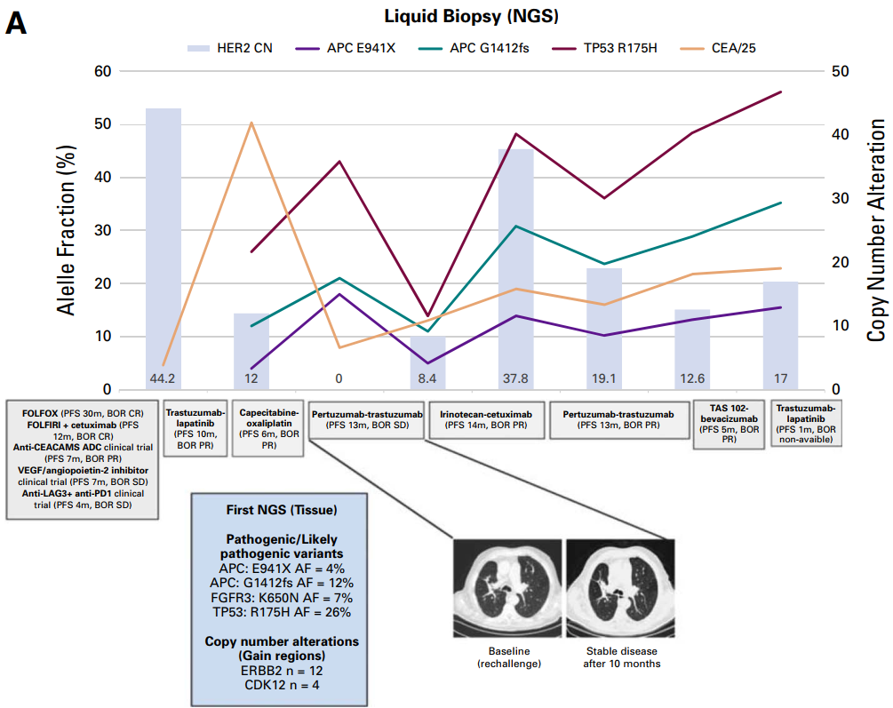
Figure 1 :Clinical course timeline and HER2 copy number evolution in case 1
Case 2: Copy number first decreases and then increases, and changing the scheme still benefits
The patient is a 53-year-old female with left colon adenocarcinoma, RAS/F wild-type, MSS, with peritoneal and ovarian metastases. He received FOLFOX + cetuximab (PFS 30 months, complete remission) and FOLFIRI + cetuximab first (PFS 10.8 months, complete remission), progressed after anti-EGFR treatment, and continued with capecitabine + oxaliplatin + bevacizumab (PFS 7 months).
During the progress, HER2 amplification was detected in the liquid biopsy (CN = 32), and the patient was treated with trastuzumab + lapatinib, which resulted in PR and lasted for 18 months.During the progress, the ctDNA showed HER2 CN decreased to 12, so the single drug of irinotecan (PFS 4 months) and XELOX (PFS 1 month) were performed, but the efficacy was not good. The liquid biopsy again showed that HER2 CN returned to 21.3, and no acquired genomic resistance mechanism was detected, so the trastuzumab + pertuzumab regimen was changed, and the clinical symptoms and tumor markers improved. It is regrettable that the patient died of pulmonary infection before the imaging evaluation, but this series of decisions itself verifies the biological rationality of ctDNA guiding the adjustment of treatment plans (Figure 2).
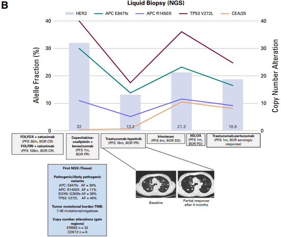
Figure 2: Clinical course timeline and HER2 copy number evolution in case 2
III. Discussion and Suggestions
These two cases show the dynamic nature of HER2-amplified mCRC: the copy number appears, disappears, and reappears with treatment pressure, suggesting that HER2-driven clones may decay and sensitivity may be restored after drug withdrawal, just like anti-EGFR-resistant clones. From the perspective of the guidelines, the NCCN guidelines for colorectal cancer have made it clear that MGPT (multi-gene panel) from tissue or blood sources can be used for molecular typing, and patients with NGS confirmed HER2 amplification are suitable for anti-HER2 treatment (Figure 3).
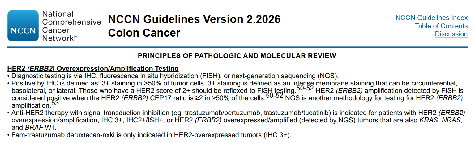
Figure 3 :The NCCN guidelines for colorectal cancer recommend HER2 overexpression/amplification testing.
However, at present there is still no clear reference for detecting HER2 amplification in ctDNAs. Whether it's more breast cancer or gastric cancer cases encountered during clinical practice, NGS methodologies are difficult to give unified consensus on determining HER2CN values and they have been used mainly as long-term dynamic monitoring tools so far.
From this, there are three tips for clinical practice:
1. HER2 amplification is a dynamic biomarker, and the idea of "measuring once and determining forever" is outdated. Copy number can be lost or duplicated; Recheck ctDNA after progress, not only to see if there are new mutations, but also to see if HER2 copy number is still there and high or not - this directly affects the next stage of treatment.
2. CTDNA liquid biopsy is a reliable alternative to timely tissue biopsy for molecular subtyping. Case 1: In the absence of an available organization, a tube of blood detected HER2 amplification (CN = 44), which opened a main line of anti-HER2 treatment that lasted for several years, and the treatment plan was changed three times, all of which were clinically beneficial.
3. There is a reciprocal relationship between HER2 and EGFR signaling axis. After HER2 amplification disappeared in case 1, the patient became sensitive to cetuximab again (PFS 14 months). HER2 amplification should not be interpreted as an "absolute resistance" marker for anti-EGFR therapy, but rather as a context-dependent variable influenced by copy number and clonal dynamics.
For the detection of HER2 amplification in ctDNA, the detection reference standard and clinical application are still being explored. With the accumulation of more clinical evidence and the standardization of technology, the information obtained from a tube of blood will bring more and more precise benefits to patients.
Reference
[1] Garcia-Pastor P, et al. Rechallenge With Anti-HER2 Therapy Guided by Liquid Biopsy in HER2-Amplified Metastatic Colorectal Cancer. JCO Precis
[2] NCCN Guidelines Version 2.2026 Colon Cancer