
2021-03-31 21:11:32

Telepathology Consultation Expert
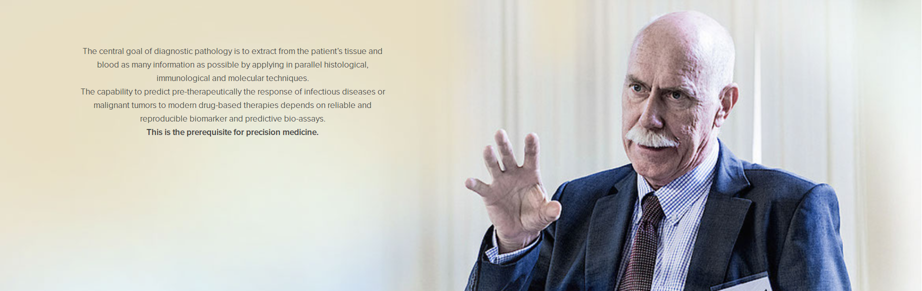
Manfred Dietel
Member of the German National Academy of Sciences Leopoldina
rofessor at the Virchow Pathology Institute, Charité – Universitätsmedizin Berlin, Germany
Case Consultation 1
Clinical Information
A 30-year-old female patient presented with a mass on the left lower lip for over 5 years. The mass was initially discovered during wisdom tooth extraction in 2016 on the buccal side of the right lower lip, measuring approximately 0.6 cm in diameter. The patient remained asymptomatic until 2020, when she requested surgical removal for cosmetic reasons.
Gross Examination
A mucosal nodule measuring 0.7 × 0.6 × 0.5 cm was resected. The surface was smooth, the cut surface showed moderate texture, and the margins appeared well-defined with a possible capsule.
Immunohistochemistry (IHC)
-
Positive Markers: SMA (small vessels+), calponin (focal+), p63 (partial+), CK5/6 (epithelial+), p40 (focal+), EMA (glandular structures+), S-100(+), CK7(+), CD117 (minor+), Ki-67 (5–10%+).
-
Negative Markers: DOG1(–), hMAM(–).
-
Special Stains: PAS with digestion (+), Alcian Blue (+).
Initial Diagnosis
-
(Left Lower Lip) A salivary gland tumor (likely a neoplasm with combined epithelial and myoepithelial differentiation, showing moderate proliferative activity).
-
Secretory carcinoma could not be entirely excluded due to S-100 positivity. Given the rarity and atypical morphology, referral to a specialized institution was recommended.
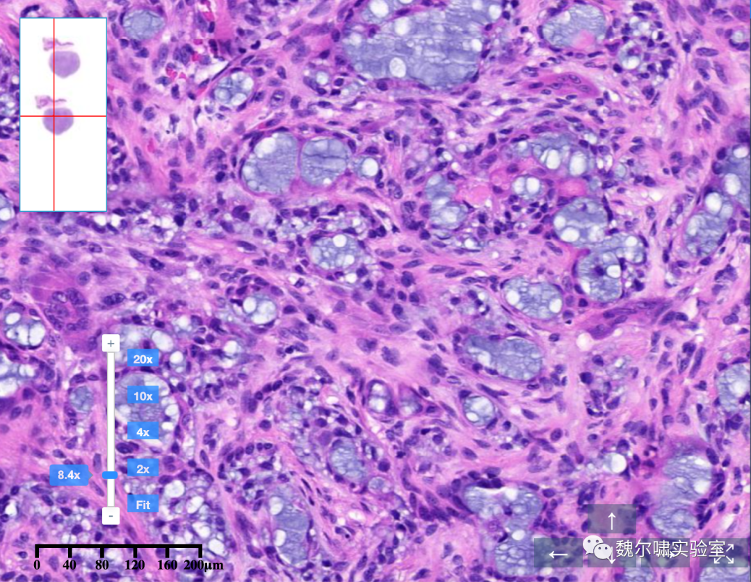
Expert Telepathology Opinion
The lesion exhibited a rounded growth pattern with orderly glandular arrangements, absent nerve invasion, and clear boundaries, consistent with a benign tumor of the minor salivary glands in the lip. Diagnosis: Myoepithelioma.
Final confirmation by Prof. Manfred Dietel (Academician, Virchow Pathology Diagnostic Center) and Dr. Pahl (Soft Tissue Pathology Specialist).
Follow-up & Summary
A diagnosis of secretory carcinoma would typically prompt aggressive surgery, causing significant physical and psychological distress. After inconclusive local consultations, the patient sought a second opinion via the Virchow International Telepathology Platform, confirming a benign myoepithelioma. No further surgery was performed, and the patient remains asymptomatic during follow-up.
Case Consultation 2
Clinical Information
A 19-year-old male presented with a localized expansile lesion in the midshaft of the tibia. MRI revealed a mass-like abnormal signal connected to the medullary cavity, with heterogeneous low signal on T1-weighted imaging and no significant enhancement post-contrast. Imaging suggested a benign lesion, but clinical correlation was advised.
Gross Examination
Specimen from the midshaft of the left tibia:
-
Soft tissue fragment: 1.0 × 0.8 × 0.3 cm.
-
Bone fragments: 0.5 × 0.3 × 0.3 cm.
Immunohistochemistry (IHC)
-
Positive Markers: P63(+), Ki-67 (10%+), VIM(+), SMA (focal+), CD68 (histiocytes+), p53 (scattered+).
-
Negative Markers: Des(–).
Initial Diagnosis
-
(Midshaft of Left Tibia) Spindle cell tumor, suspicious for osteosarcoma. Referral to a specialized center was recommended.
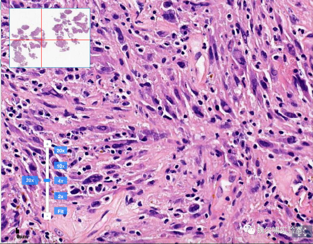
Expert Telepathology Opinion
Histology revealed invasive growth patterns:
-
Medullary invasion: Replacement of medullary space with tumor cells surrounding native trabeculae and Haversian systems.
-
Cortical destruction with soft tissue infiltration.
-
Cellular Features: Marked atypia (pleomorphic, hyperchromatic nuclei) with diverse morphologies (epithelioid, plasmacytoid, spindle, small round, clear cells, and giant tumor cells).
Final Diagnosis: Conventional Osteosarcoma (High-Grade).
Additional IHC recommended: S100, osteocalcin, CD99, SMA (typically at least focal positivity); CD31 (–).
Follow-up & Summary
Osteosarcoma diagnosis requires integration of clinical, imaging, and histologic data. Despite imaging initially suggesting benign features, the patient received a definitive diagnosis via Virchow International Telepathology. He underwent a second surgery in Shanghai and is currently on his 8th cycle of osteosarcoma-specific chemotherapy, with no recurrence or metastasis observed.
Telepathology Consultation Services
Pathology diagnosis is pivotal in clinical practice—accurate diagnosis dictates appropriate treatment, while errors may lead to catastrophic outcomes. Pathologists are often likened to "judges" in medicine, delivering the final "verdict" on a patient’s condition.
However, becoming a qualified "judge" is exceptionally challenging. Unlike other specialties, pathologists in China are not subspecialized. A general pathologist must diagnose over 10,000 diseases across all organs and tissues, requiring mastery of gross, histomorphologic, immunophenotypic, and molecular genetic features. This is particularly daunting in primary care settings, where rare or complex cases often lack definitive diagnoses.
Pathology Consultation has thus emerged as a critical solution. Patients and clinicians seek second opinions from renowned experts at large academic centers, where pathologists possess broader experience and exposure to diverse cases.
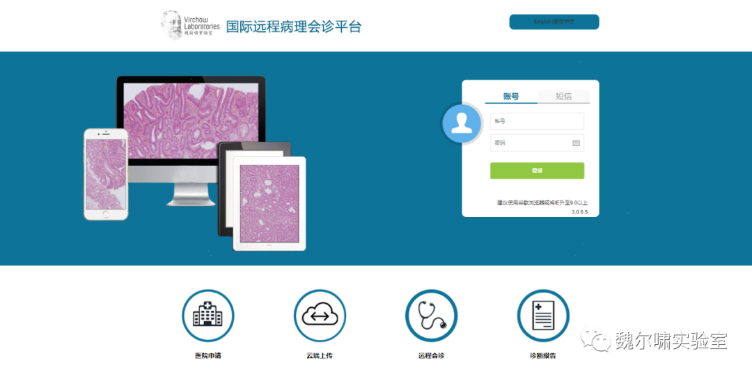
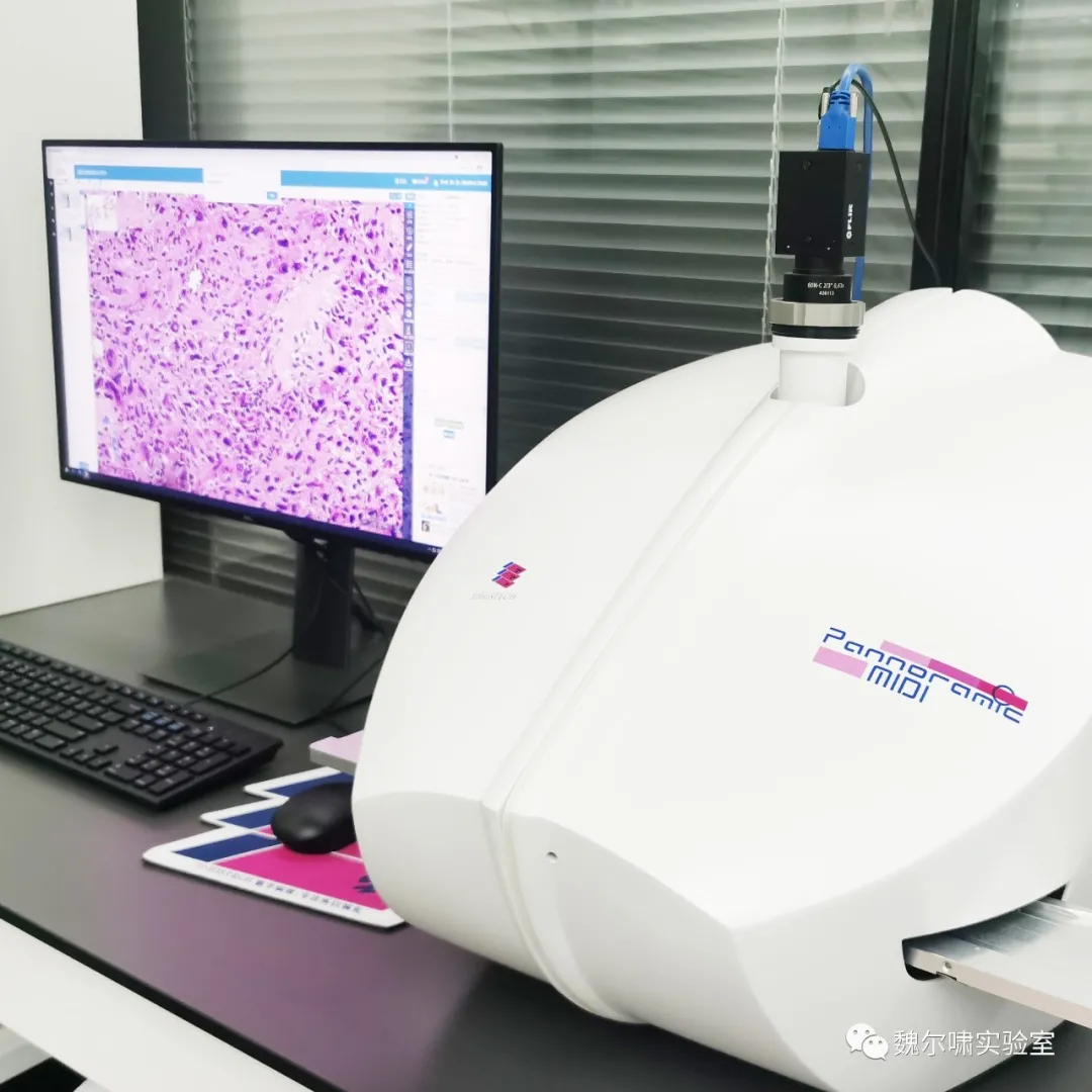
Virchow Laboratory addresses this need through its Telepathology Consultation Platform, enabling patients to access top-tier pathology expertise globally without leaving their local communities.
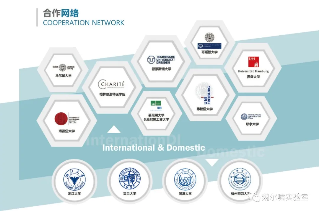
Virchow Laboratory’s Telepathology Network
We have established a Global Telepathology Network through internet-based systems, collaborating with leading academic institutions and pathologists worldwide. Key deployments include Shanghai, Hangzhou, Berlin, and multiple German pathology institutes. The Charité – Universitätsmedizin Berlin Pathology Institute serves as the central hub for international case distribution.
Current Network:
-
22 academic institutions in Germany.
-
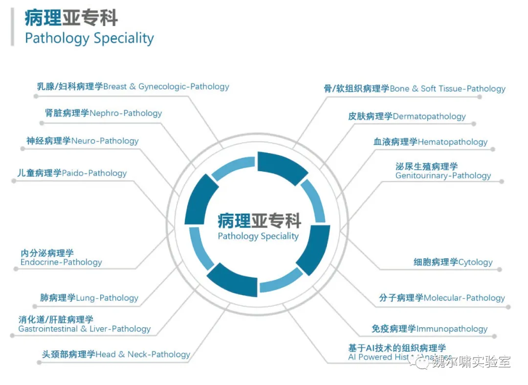
16 subspecialty divisions.
-
200+ pathologists.
-
AI-powered workflows for rapid, high-quality diagnostics.
Innovative Technologies:
-
High-resolution virtual microscopy via advanced slide-scanning and algorithms.
-
Integrated molecular profiling for comprehensive assessments.
Services Offered:
-
Comprehensive Pathology Services: Routine diagnostics, intraoperative frozen sections, and expert consultations.
-
Second Opinions: For pathologists, clinicians, or patients.
-
AI-Driven Biomarker Quantification: Ki67, ER, PR, HER2, ALK mutations, and expanding panels.
-
Integrated Data Analysis: Combining histomorphology, imaging, and molecular data to guide personalized treatment.
This platform revolutionizes pathology by bridging global expertise, cutting-edge technology, and patient-centered care.